
Patel Nagar, Hargobind Nagar, Phagwara, Punjab 144401
Email: mitraeyehospital@gmail.com
Phone: 01824-356072
TPA: 01824-357551
Reception: 98888-18840
Appointment: 95011-16997
Monday to Saturday – 10am to 6pm
Vitreoretinal surgery in Jalandhar, Phagwara, Punjab, Mitra Eye Hospital under the supervision of Dr. Harinder Mitra is fully equipped with state-of-the-art Vitreo Retinal Surgical Treatment. This surgical approach is performed under various issues linked to retinal problems. The 3D Genuinity visualization system helps the surgeon to perform the heads-up vitreoretinal surgery. Vitreoretinal surgery Cost in Punjab starts from Rs 20,000 and it can range high depending on your condition.
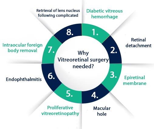
Vitreoretinal Eye Surgery is performed under the group of latest procedures to treat the inside of the eyes with conventional or laser surgical instruments. Under the supervision of Dr. Harinder Mitra, Dr. Akshay Mitra and his expert team will perform this delicate surgery on the part where there is gel-like vitreous and retina (light-sensitive membrane) are found. This approach is beneficial for different eye conditions which are linked to age-related macular degeneration, macular hole, detached retina, CMV retinitis, epiretinal membrane, and diabetic vitreous hemorrhage.
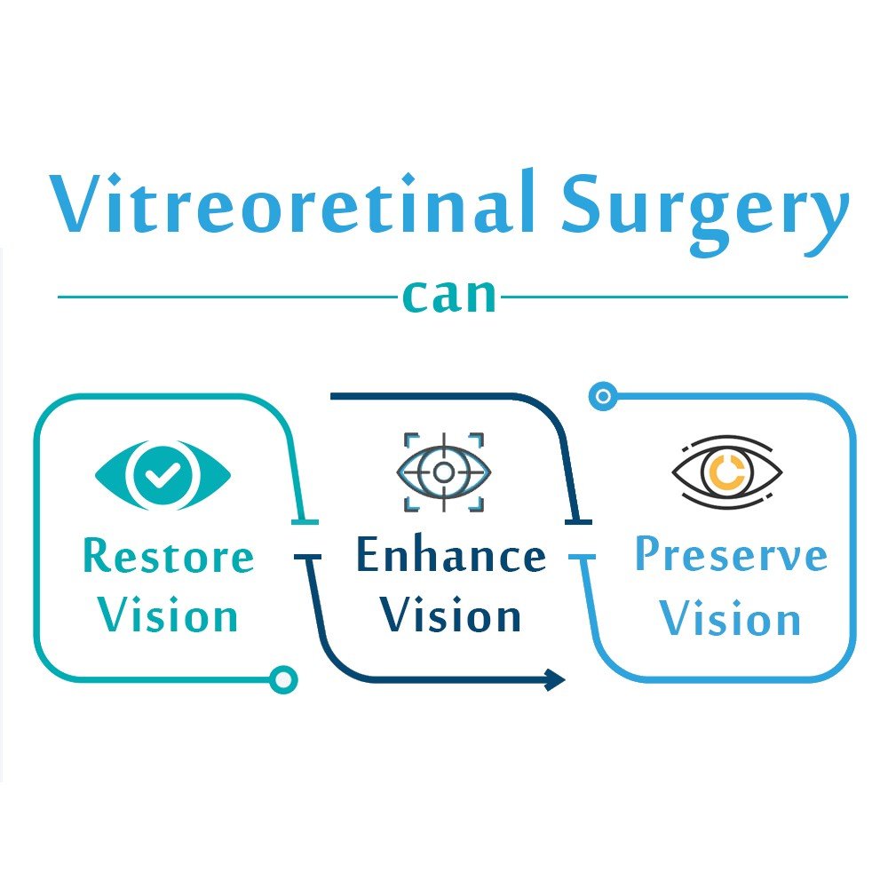
| Do you know what a vitreoretinal surgical procedure can do? | ||
| Restore Vision | Preserve Vision | Enhance Vision |
Like with any other surgery, the doctor will make sure that you are a good candidate for the procedure. MITRA EYE HOSPITAL ophthalmologists will do a thorough eye examination and go through your health history to make sure you are an ideal candidate for the treatment. Our team of eye specialists closely evaluates the patient’s condition and then suggests to them the best possible treatment plan.
Symptoms reduced by Contoura Vision
Get yourself freedom from many of the symptoms linked with LASIK and SMILE.
Medical institutes have been continuously working on researching innovative techniques for better results and outcomes. Contoura Vision is an outcome of one such research and effort. Ever since 1990, the oculists have been experimenting with the processes that can best result in corneal surgery Contoura vision satisfies their search. It is a wavefront analysis that maps the entire eye of the patient.
The principle of Contoura vision is a detailed study of the topography of the eye. It can study 2,000 cornea points precisely, unlike LASIK wave technology that can reach only 200 corneal points. Capturing many cornea points adds to the efficiency and precision that Contoura vision results bring. It drastically enhances the vision, clarity, sharpness, and all this involves minimum at the side of the patient. The procedure needs no incisions, scars, or bandages.
It causes minimum pain and also requires no hospitalization in many cases. It is like a walk-in, get the procedure done, and walk out from the clinic feeling near to fit. Whatever may be the advantages of Contoura vision, it has its restrictions in terms of recommendations. It is your oculist who decides and recommends Contoura vision depending on many medical and health perspectives that vary from individual to individual. Mitra Eye hospital has a team of expert oculists who take into consideration every detail of the patient before they opt for any eye surgeries.
How does the Vitrectomy procedure work?
During the surgery, the gel-like substance or vitreous humor is removed. Undergoing this approach will solve the vision issues when the foreign matter affects the pristine area which is the inner part of the eyes. One of the foreign substances in the blood that can occur due to diabetic vitreous hemorrhage.
Once the vitreous humor is taken away and the entire area is cleaned. After that, the saline liquid is injected to replace it with the vitreous humor
Once the surgeon removes the vitreous humor and clears the area, he or she usually injects a saline liquid to replace the vitreous humor that ordinarily fills up the inner chambers of the eye which fills in the inner part of the eyes.
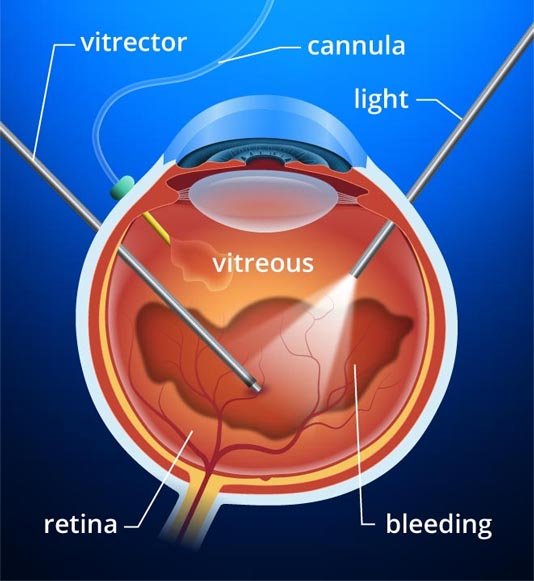
In most cases, the patient needs general anesthesia and sometimes local anesthesia is used. The surgeon will create tiny incisions (around 3) to create the eye-opening and through the opening instruments are added. Inside the incisions, the doctor will place the instruments which include:
Epiretinal membrane (ERM) is also referred to as macular pucker and cellophane retinopathy includes membrane growth which is the same as that of scar tissue around the macula. Many times it happens that the macula, the sensitive part of the retina responsible for the visual activity and color vision, encounters growth of a scar across the retina, causing central vision distortion. The solution to the problem is Membranectomy, also known as macular pucker or cellophane retinopathy. After completing the process of vitrectomy, the surgeon making use of precise, sharp forceps will peel away the membrane from the retina. Tiny sutures seal the incisions that do not require removal. The recovery rate from the membrane to my is high in many cases, yet exceptions prevail. It does not carry many risks with it. However, not following the precautionary measures can lead to infection and increase the time of recovery.
Reasons to get membranectomy | |
| Epiretinal Membrane is present | Problem of Vision Distortion or reduced vision due to ERM. |
Initially, the procedure starts with a vitrectomy. The surgeon uses the fine forceps under high magnification to gasp and feel the membrane from the retina. The membrane is removed by the diamond-dusted instruments. Our surgeon performs the surgery with utmost precision to make sure eyes are not affected.
Following retinal detachment, the patient will have the most common complication named Proliferative vitreoretinopathy (PVR). The doctor will diagnose it and then perform the surgery.
With time the retinal tissue becomes rigid, hard, and immobile increasing visionary and treatment problems. Ironically this problem has a reverse relation to the treatment. In other words, during the developmental stage, the retina is compliant, and PVR at this stage is not possible as the problem lacks clarity.
The retina is neither in its exact form nor completely deformed in shape, location. Over the period, the retina becomes detached, rigid taking the white, fibrotic appearance increasing the chances of removal. Vitrectomy follows the filling of gases and liquids into the eye to help keep the retina reattached to the outer walls of the eye.
The process many times also calls in need for sclera. Artificial material is inserted into the eye to maintain the pressure that helps in keeping the retina attached to the wall. The results of the process enable Ambulatory vision, but the precise and sharp clarity in vision remains far from reality.
50% of the patient will have a vision but that is to see the objects within a close range. Although, the reading vision will be low.
Created by Fatewise Digital Solutions
Copyright © 2024. All rights reserved.